

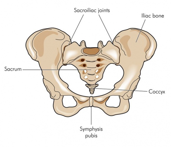
The sacrum and coccyx are two anatomical structures located near the bottom of your vertebral spinal column, below the fifth lumbar vertebra (L5). The sacrum, sometimes called the sacral spine (abbreviated S1), is a large, flat triangular-shaped bone located below L5 and in between your hip bones. Below the sacrum is the coccyx, commonly known as the tailbone.
Individually, the sacrum and coccyx are composed of smaller bones that fuse (grow into a solid bone mass) together by age 30. The sacrum is made up of 5 fused vertebrae, and 3 to 5 small bones fuse to create the coccyx. Both structures are weight-bearing and integral to functions such as walking, standing and sitting.
General MRI Preparation
Each patient is provided their own private locked dressing room which has an area to hang their clothing. We ask that patients do not enter the scanning room with jewelry, watches, keys, coins or other metal objects. Credit card and ATM card magnetic codes will be erased by the MRI’s magnetic field if brought into the room.
Patient position
The radiograph is performed with the patient in a lateral recumbent position
- the patient can be either on the left or right lateral recumbent position, depending on which is more comfortable
- flex the knees
- a cushion under the waist can aid patient comfort
- ensure patient is in a true lateral position
Image technical evaluation
- the entire sacrum and coccyx should be visible from L5/ S1- terminal coccyx
- no patient rotation as demonstrated by superimposition of the greater sciatic notches and femoral heads
- adequate penetration should clearly demonstrate the sacrum/coccyx region